ARV-825 targeted BRD4 degradation suppresses nuclear PD-L1 RelB IL-6 signaling in breast cancer
Copy code to add this image to your page
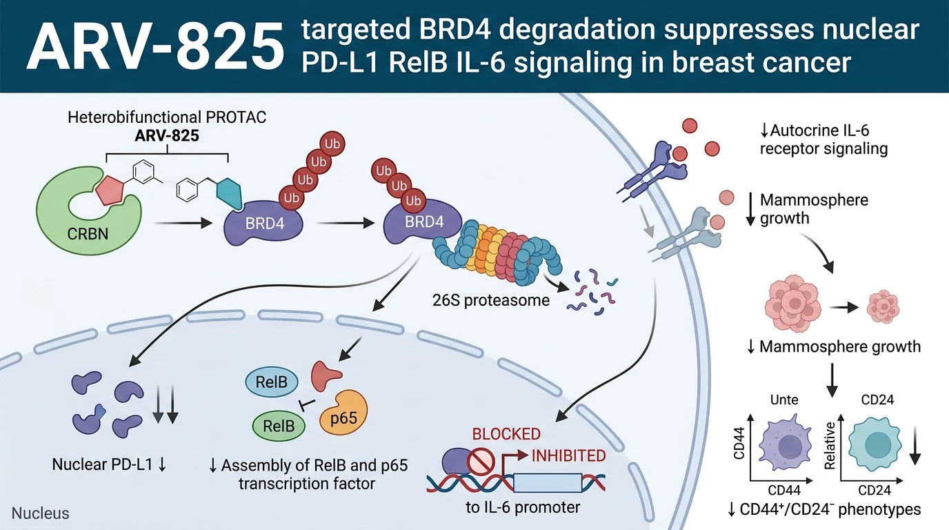
Bromodomain-containing protein 4 (BRD4), a key member of the bromodomain and extra-terminal (BET) protein family, functions as an epigenetic reader that recognizes hyperacetylated lysine residues on histone tails. Through its dual bromodomains (BD1 and BD2), BRD4 anchors to active enhancers and super-enhancers, recruiting positive transcription elongation factor b (P-TEFb) to stimulate RNA polymerase II-dependent transcript elongation. In breast cancer stem cell (BCSC) subpopulations, BRD4 expression levels are markedly elevated compared to non-stem bulk tumor populations. This differential expression sustains transcriptional networks governing self-renewal, metabolic plasticity, and immune evasion. Targeted chemical clearance of BRD4 using the small-molecule PROTAC ARV-825 provides an effective modality to dismantle these driver transcriptional complexes. Synthesized as a heterobifunctional chimera linking a triazolo-zeranol derivative to a cereblon (CRBN) E3 ubiquitin ligase ligand, ARV-825 brings BRD4 into spatial proximity with the E3 ligase machinery, initiating rapid polyubiquitination and subsequent 26S proteasomal degradation of the target protein.
In cultured breast cancer cell lines and patient-derived cell models, exposure to nanomolar concentrations of ARV-825 achieves near-complete depletion of BRD4 within hours. This catalytic turnover contrasts sharply with traditional small-molecule occupancy-based bromodomain inhibitors, which often trigger compensatory BRD4 protein accumulation. Disruption of BRD4-dependent transcription significantly impairs the self-renewal capacity of BCSCs. In vitro anchorage-independent mammosphere assays show marked suppression of both primary and secondary sphere counts following exposure to the degrader. Flow cytometric evaluations confirm a concurrent reduction in the proportion of CD44+/CD24- stem-like cells, alongside diminished aldehyde dehydrogenase 1 (ALDH1) enzymatic activity, an established functional marker of stemness.
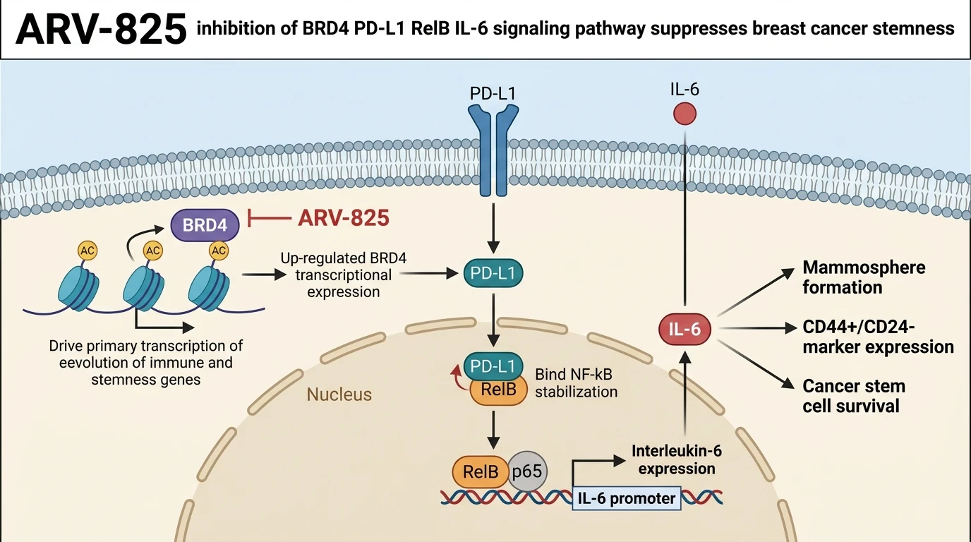
At the molecular level, BRD4 binds promoter and enhancer elements of the CD274 gene, which encodes programmed cell death 1 ligand 1 (PD-L1). While membrane-bound PD-L1 acts primarily as an immune checkpoint ligand that induces T cell exhaustion, an intracellular subpopulation translocates to the nucleus. ARV-825-mediated destruction of BRD4 selectively abolishes CD274 transcription, leading to rapid exhaustion of both cytosolic and nuclear PD-L1 pools. In the nuclear compartment, PD-L1 operates in a non-canonical capacity, modulating chromatin access and acting as a transcriptional co-activator for specific target genes, including RelB.
RelB, an essential mediator of the non-canonical NF-kB pathway, exhibits decreased transcript and protein abundance following PD-L1 depletion. Nuclear PD-L1 physically interacts with promoter regions of the RelB locus to sustain its active transcription. Upon synthesis, RelB forms functional heterodimeric transcription factor complexes with p65 (RelA). The assembled RelB/p65 complex translocates into the nucleus and binds specific kappaB motif sequences located within the interleukin-6 (IL-6) promoter, initiating robust transcription of IL-6 mRNA. Secreted IL-6 subsequently binds to its cognate receptor alpha (IL-6R) and signal transducer gp130, activating Janus kinase (JAK) signaling and downstream STAT3 phosphorylation. This autocrine loop directly reinforces BCSC survival and sphere-forming capacity.
Pharmacological intervention with ARV-825 dismantles this cascading signaling axis at its origin. Loss of BRD4 leads to immediate collapse of nuclear PD-L1 levels, preventing RelB transcription, halting RelB/p65 complex assembly, and arresting IL-6 gene expression. Experimental evidence confirms that exogenous supplementation with recombinant IL-6 partially restores mammosphere formation in ARV-825-treated cells, confirming the central functional role of IL-6 down-regulation in mediating anti-stemness responses. Furthermore, suppression of PD-L1 and IL-6 within tumor tissue decreases local immunosuppressive signaling, enhancing cytotoxic T cell recognition in co-culture models. In addition to cell-autonomous stemness attenuation, the systemic effects of targeted BRD4 degradation touch upon the broader tumor microenvironment. Reduced IL-6 secretion limits paracrine recruitment of myeloid-derived suppressor cells and tumor-associated macrophages in preclinical models. This dual activity—simultaneously targeting intrinsic stem cell self-renewal and extrinsic immunosuppressive cytokine networks—underlines the multi-layered pharmacodynamics of PROTAC-mediated BRD4 destruction.
Targeted BRD4 Proteolysis Attenuates Nuclear PD-L1 Mediated Stemness Cascades
Copy code to add this image to your page
ARV-825 is a PROTAC degrader targeting BRD4 for proteasomal degradation. In breast cancer stem cells, BRD4 drives transcription of PD-L1, which translocates to the nucleus to regulate RelB expression. The resulting RelB/p65 complex promotes IL-6 transcription to maintain stemness. ARV-825-mediated BRD4 degradation depletes nuclear PD-L1, suppresses RelB and IL-6 expression, inhibits mammosphere formation, reduces CD44+/CD24- and ALDH1 markers, and suppresses stemness while enhancing antitumor immune responses.
BRD4 Proteolysis Suppresses BCSC Mammosphere Formation
Exposure of breast cancer cell cultures to ARV-825 induces rapid, concentration-dependent degradation of BRD4 protein. Target depletion occurs within four hours in vitro. Proteasomal degradation was completely blocked by pre-incubation with the proteasome inhibitor MG132; this confirms E3 ligase-dependent target turnover. Functional assays demonstrate that the compound severely restricts primary and secondary mammosphere formation in triple-negative breast cancer lines. The total count of CD44+/CD24- stem-like cells decreased by over sixty percent relative to vehicle control. ALDH1 enzymatic activity fell in parallel. Downstream phosphorylation of STAT3 decreased markedly within twelve hours. These observations confirm that continuous epigenetic maintenance by BRD4 is mandatory for BCSC self-renewal.
Nuclear PD-L1 RelB Direct Transcription Drives Interleukin-6 Expression
Mechanistic analysis revealed an unexpected non-canonical role for PD-L1 in BCSC maintenance. Depletion of BRD4 using the heterobifunctional degrader reduced total CD274 mRNA levels by seventy-five percent in patient-derived stem cells. Subcellular fractionation demonstrated a corresponding loss of nuclear-localized PD-L1 protein. Nuclear PD-L1 directly bound chromatin at the RelB promoter region—a physical interaction mapped via chromatin immunoprecipitation assays. Diminished nuclear PD-L1 impaired RelB mRNA expression; cytosolic RelB protein levels dropped sharply. Consequently, formation of the transcriptionally active RelB/p65 heterodimer ceased. Electrophoretic mobility shift assays showed complete loss of RelB/p65 binding to the IL-6 promoter kappaB consensus sequence. Secreted IL-6 concentration in culture media dropped below detectable limits within twenty-four hours.
Targeted BRD4 Disruption of Epigenetic Signaling Restores Antitumor Immunity
Interruption of the signaling axis produces distinct biological consequences in co-culture systems containing peripheral blood mononuclear cells. Suppressed IL-6 secretion reduced BCSC-mediated induction of immunosuppressive M2 macrophage phenotypes. Concurrently, down-regulation of surface PD-L1 on tumor cells restored CD8+ cytotoxic T lymphocyte proliferation and granzyme B secretion. In immunocompetent mouse models, administration of ARV-825 led to tumor regression without significant systemic toxicity. Tumor re-inoculation assays demonstrated a three-fold increase in the minimum cell number required for graft establishment. These findings indicate that targeting BRD4 destabilizes intrinsic stemness programs while simultaneously reactivating local antitumor immune responses.