ANA-12 Trk receptor antagonist suppresses hippocampal neurodevelopment and mitochondrial bioenergetics
Copy code to add this image to your page
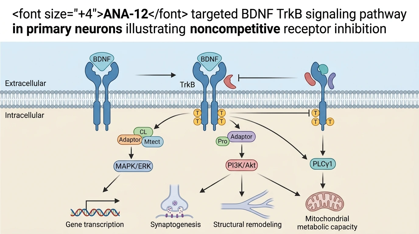
The Tropomyosin-related kinase B (TrkB) receptor serves as a primary conductor of activity-dependent plasticity, dendritic arborization, and structural synaptogenesis across mammalian central neural networks. Under baseline physiological conditions, binding of the homodimeric neurotrophin brain-derived neurotrophic factor (BDNF) to the extracellular domain of TrkB triggers receptor dimerization and transphosphorylation of cytoplasmic tyrosine residues—specifically Tyr515 and Tyr816. These phosphorylated residues act as high-affinity docking sites for adaptor proteins including Shc, Grb2, and PLCγ1, initiating downstream cascade branches through the Ras/MAPK, PI3K/Akt, and PLCγ1/IP3/DAG signaling axes. Pharmacological dissection of this neurotrophic cascade necessitates high-precision small-molecule tools capable of discriminating TrkB from closely related receptor tyrosine kinases such as TrkA and TrkC. Within this mechanistic context, the synthetic low-molecular-weight ligand ANA-12 Trk receptor antagonist functions as a selective, non-competitive inhibitor that engages allosteric domains on TrkB, preventing BDNF-mediated receptor autophosphorylation and downstream signal transduction without altering basal receptor expression levels or inducing intrinsic receptor internalization.
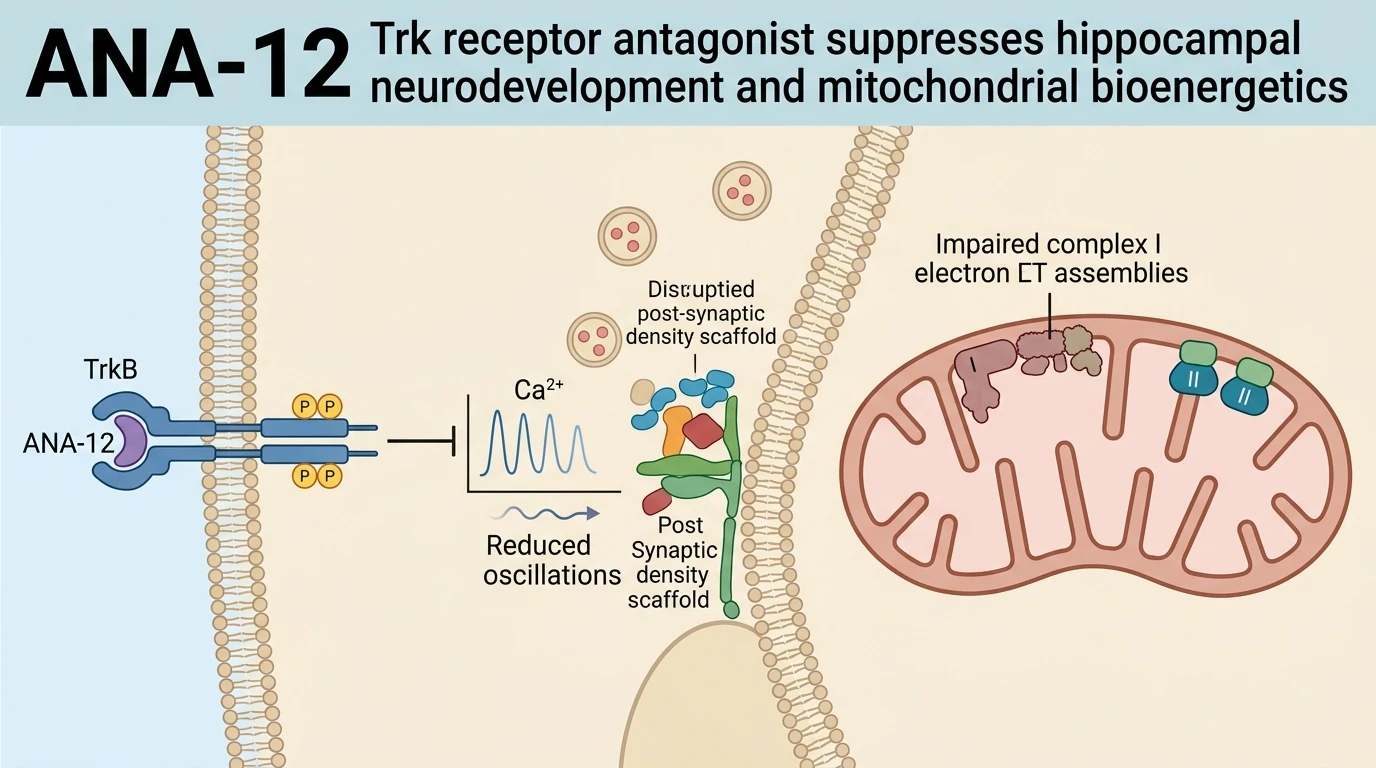
To evaluate the necessity of constitutive TrkB signaling during network formation, primary hippocampal neural-glial co-cultures were subjected to sustained pharmacological modulation during critical developmental windows in vitro. Multi-electrode array (MEA) electrophysiology revealed that chronic administration of 1 µM ANA-12 significantly reduced spontaneous bioelectrical activity. Mean firing rates declined rapidly, accompanied by a marked reduction in network-wide burst frequency and duration. In untreated or vehicle-controlled assemblies, cultured primary neurons spontaneously organize into scale-free functional topologies anchored by highly connected 'hub' neurons that coordinate collective firing patterns. Treatment with ANA-12 disrupted this functional topology; cross-correlation analyses demonstrated a significant degradation of functional connectivity vectors and a complete arrest of functional hub node maturation.
Complementary real-time calcium imaging utilizing fluorescent indicators revealed corresponding alterations in intracellular second-messenger dynamics. Chronic TrkB blockade by ANA-12 decreased both the amplitude and temporal synchronization of spontaneous somatic calcium oscillations across multi-neuronal fields. Under control conditions, calcium transients propagated as highly ordered inter-cellular waves through astrocyte-neuron networks; however, ANA-12 administration uncoupled these oscillatory dynamics, reducing baseline cytosolic calcium flux and inhibiting activity-dependent calcium influx through L-type voltage-gated calcium channels and NMDA receptors.
Ultrastructural analysis by transmission electron microscopy provided direct physical evidence for the functional deficits observed during electrophysiological recordings. Neuronal processes treated chronically with 1 µM ANA-12 exhibited delayed synaptogenesis characterized by decreased numerical synaptic density, shortened active zone lengths, and reduced electron density (osmiophility) within the post-synaptic density (PSD) protein matrix. Beyond structural synaptic deficits, ANA-12 exposure induced significant subcellular organelle pathology, specifically targeting mitochondrial cytoarchitecture. Micrographs revealed mitochondrial swelling, disrupted cristae alignment, and matrix vacuolation within pre- and post-synaptic terminals. Spectrophotometric enzymatic assays demonstrated that these structural defects coincided with a selective decline in mitochondrial respiratory chain complex I (NADH:ubiquinone oxidoreductase) activity, while complex III and IV activities remained largely unaffected.
Exogenous application of recombinant BDNF elicited reciprocal effects, driving hyper-synchronous network bursting, elevated calcium transient amplitudes, heightened PSD osmiophility, and a selective increase in mitochondrial respiratory complex II (succinate dehydrogenase) activity. Significantly, co-incubation of BDNF with 1 µM of ANA-12 completely prevented all BDNF-induced electrophysiological, structural, and metabolic enhancements. These empirical observations demonstrate that TrkB catalytic activation is indispensably required for structural synaptogenesis, functional network connectivity, and mitochondrial metabolic maintenance in developing hippocampal networks under in vitro culture conditions.
Pharmacological Evaluation of TrkB Inhibition in Hippocampal Networks
Copy code to add this image to your page
ANA-12 acts as a selective, non-competitive antagonist of the Tropomyosin-related kinase B (TrkB) receptor. By engaging allosteric binding sites on the TrkB extracellular/transmembrane interface, ANA-12 prevents BDNF-mediated receptor dimerization and autophosphorylation. This blockade inhibits downstream activation of the Ras/MAPK, PI3K/Akt, and PLCγ1 pathways. Consequently, ANA-12 suppresses spontaneous bioelectrical firing, disrupts calcium wave synchronization, arrests functional hub node maturation, delays structural synaptogenesis, and impairs mitochondrial respiratory chain complex I activity in primary hippocampal neural-glial networks.
Electrophysiological Desynchronization and Functional Network Connectivity Suppression
Long-term recording across multi-electrode arrays demonstrated that TrkB inhibition alters intrinsic network biophysics. Exposure to 1 µM ANA-12 reduced spontaneous action potential discharge rates within 48 hours. Mean burst frequency fell by over 60% compared to vehicle controls. Furthermore, functional connectivity matrices derived from cross-correlation algorithms revealed a loss of scale-free network topology. The formation of highly connected hub neurons failed entirely under persistent ANA-12 Trk receptor antagonist exposure. Spontaneous somatic calcium oscillations lost temporal coherence across neighboring cells. Cytosolic calcium wave propagation velocity decreased significantly. These data indicate that constitutive TrkB receptor activation provides necessary signaling tone to drive early bioelectrical synchronization.
Ultrastructural Synaptic Retardation and Mitochondrial Complex I Impairment
Transmission electron microscopy confirmed distinct subcellular structural lesions following non-competitive TrkB receptor blockade. Synaptic density dropped markedly in primary hippocampal cultures treated with 1 µM ANA-12. Active zone lengths were diminished. Post-synaptic density osmiophility—a surrogate for dense protein scaffold assembly—was significantly attenuated. Mitochondria exhibited severe ultrastructural damage characterized by outer membrane distension, cristae fragmentation, and matrix rarefaction. Spectrophotometric micro-assays showed a targeted 35% reduction in mitochondrial respiratory complex I activity. Exogenous BDNF failed to restore complex I activity or synaptic density when co-administered with the selective ANA-12 Trk receptor antagonist. Thus, TrkB signaling directly regulates structural synaptogenesis and mitochondrial respiratory chain integrity in primary neural networks.