β-Aminopropionitrile βAminopropionitrile lysyl-oxidase inhibition destabilizes aortic medial elastic lamellae
Copy code to add this image to your page
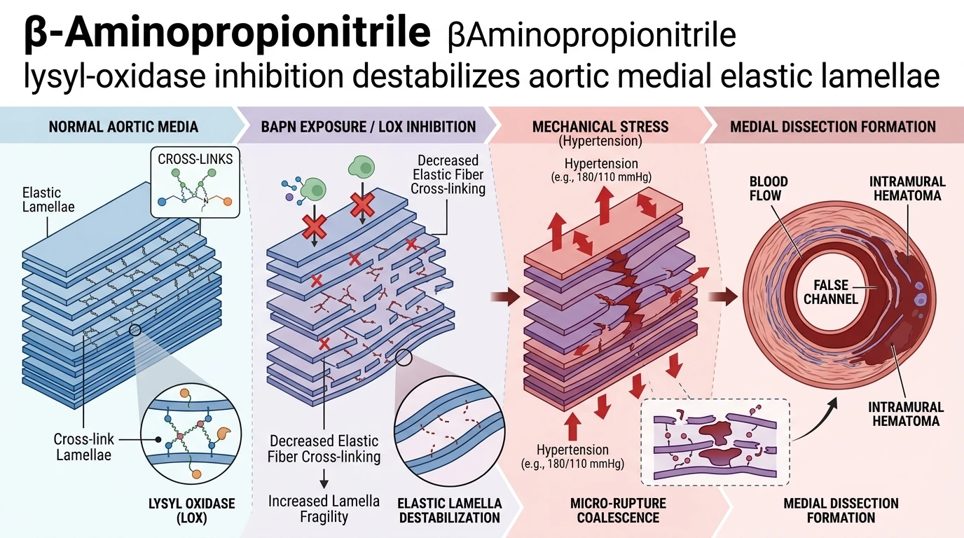
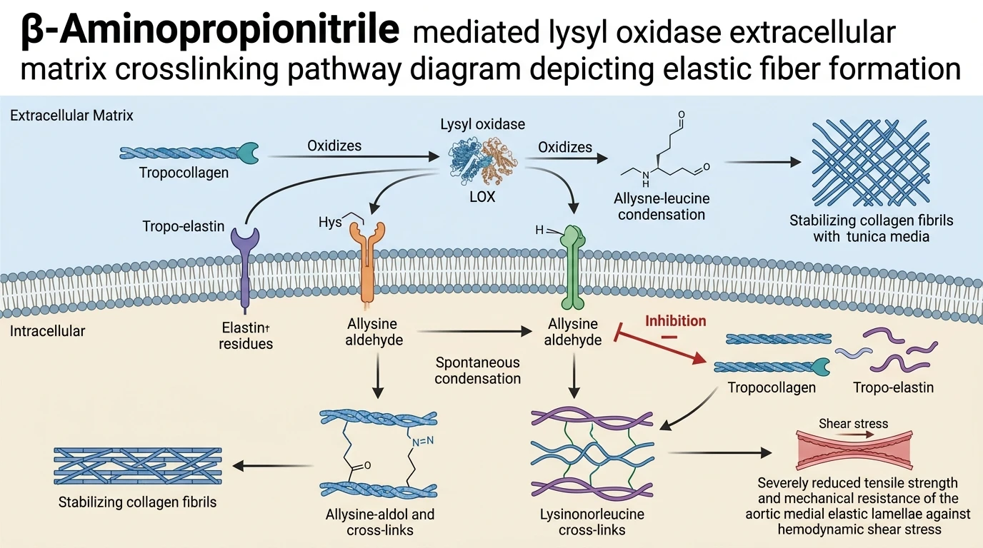
Aortic dissection remains a lethal cardiovascular pathology characterized by sudden tearing of the aortic tunic media. The structural stability of the aortic wall relies on the integrity of the extracellular matrix (ECM), particularly concentric elastic lamellae interspersed with smooth muscle cells and collagen networks. Lysyl-oxidase (LOX) is a copper-dependent amine oxidase that plays an indispensable role in maintaining arterial tensile strength. LOX catalyzes the oxidative deamination of lysine and hydroxylysine residues in tropocollagen and tropo-elastin precursors. This enzymatic modification generates reactive allysine aldehydes that spontaneously condense to form covalent interchain cross-links, such as desmosine, isodesmosine, and lysinonorleucine. These covalent bonds consolidate monomeric tropocollagen and tropo-elastin into mature, insoluble collagen fibrils and elastic fibers capable of withstanding repetitive pulsatile blood pressure.
Pharmacological inhibition of lysyl-oxidase by β-Aminopropionitrile interrupts this maturation cascade. β-Aminopropionitrile functions as an irreversible suicide substrate that covalently modifies the active site of LOX. Consequently, newly synthesized tropo-elastin and tropocollagen monomers fail to form functional cross-links within the medial matrix. While pre-existing cross-linked fibers remain temporarily intact, endogenous matrix turnover gradually depletes mature fibers. Over time, the newly deposited, uncrosslinked matrix components lack structural cohesion, inducing profound mechanical instability and structural fragility within the medial elastic lamellae.
When LOX inhibition by β-Aminopropionitrile is paired with Angiotensin II (AngII)-induced hypertension, the aortic wall is subjected to severe mechanical strain. AngII elevates systemic blood pressure through AT1 receptor-mediated vasoconstriction and fluid retention, driving circumferential wall stress. Under elevated pressure, the biochemically compromised, uncrosslinked elastic lamellae cannot effectively dissipate hemodynamic force. This dynamic synergy between LOX inhibition and hypertensive load precipitates catastrophic structural breakdown within the aortic wall.
Recent spatial studies utilizing synchrotron-based 3D micro-tomography analysis have clarified the exact spatial evolution of aortic medial ruptures at individual elastic lamella resolution in mouse models receiving β-Aminopropionitrile and AngII. Propagation-based phase-contrast synchrotron imaging provides unprecedented isotropic spatial resolution, permitting non-destructive visual dissection of individual medial layers across the thoraco-abdominal and ascending aorta. Within 3 days of co-administration, localized single-layer (L1) micro-ruptures and initial multi-layer (ML) elastic lamellar tears emerge within the tunica media. These early focal ruptures occur primarily at points of maximum mechanical stress without immediate catastrophic vessel collapse.
By day 14 of continuous administration, these localized micro-ruptures undergo dramatic volumetric expansion and continuous spatial coalescence. Ultra-high-resolution three-dimensional reconstructions reveal that isolated focal tears bridge adjacent elastic lamellae, converting small micro-cracks into extensive contiguous medial tears. In the thoraco-abdominal aorta, this coalescence manifests as complete medial dissections, driven by pressurized blood forcing its way into torn lamellar planes to form functional false channels and large intramural hematomas. Conversely, in the ascending aorta, structural failure predominantly presents as interlamellar hematomas, where blood accumulates between disrupted lamellar sheets without forming a continuous secondary conduit.
These high-resolution imaging observations confirm that aortic dissection is not an instantaneous single-point failure. Instead, it proceeds through an ordered micro-structural cascade: biochemical destabilization of ECM cross-linking via LOX inhibition, initiation of discrete single-lamella micro-ruptures under hypertensive stress, and subsequent 3D coalescence into macro-scale medial dissections. Mechanistic characterization of this progression highlights the imperative of maintaining covalent elastic matrix cross-linking to preserve vessel resistance against hemodynamic mechanical strain.
Structural Mechanics and Medial Rupture Progression in Aortic Dissection
Copy code to add this image to your page
Irreversible inhibition of lysyl-oxidase by β-Aminopropionitrile prevents covalent cross-linking of tropo-elastin and tropocollagen monomers. This biochemical blockade impairs elastic fiber maturation and destabilizes the aortic medial framework. Under Angiotensin II-induced hypertension, uncrosslinked elastic lamellae undergo rapid mechanical shear failure. Micro-ruptures initiate within individual medial lamellae (L1) and coalesce into multi-layer (ML) tears over 14 days. Medial dissection ensues as high-pressure arterial flow enters ruptured elastic layers, forming false channels and extensive intramural hematomas across the aortic architecture.
Synchrotron Imaging Identifies Early Elastic Lamellar Micro-Ruptures
Phase-contrast synchrotron tomography mapped initial medial lesions at single-lamella resolution. Administration of βAminopropionitrile impaired lysyl-oxidase activity in C57BL/6J mice. Within three days, isolated micro-ruptures appeared in single elastic lamellae (L1). These early focal defects did not instantly compromise total arterial wall boundary conditions. Wall stress increased locally at micro-rupture margins. Micro-rupture sites exhibited elevated strain concentrations under elevated systolic pressure; structural strain concentrated along adjacent unruptured elastic fibers.
Coalescence of Micro-Ruptures Drives Medial False Channel Formation
Micro-ruptures expanded spatially over 14 days of sustained hypertension. Volumetric analysis demonstrated that discrete single-layer tears coalesced into multi-layer (ML) disruptions. In animals receiving βAminopropionitrile and Angiotensin II, continuous mechanical cleavage separated adjacent medial sheets. Blood penetrated these torn lamellar spaces. Pressurized delamination created false channels in the thoraco-abdominal aorta; intramural hematomas expanded within the outer third of the tunica media.
Hemodynamic Stress Amplifies Multilayer Elastic Fiber Fragmentation
Regional biomechanical differences shaped distinct dissection phenotypes across the aortic arch. Ascending aortic sections developed extensive interlamellar hematomas rather than primary false conduits. Uncrosslinked tropo-elastin matrix failed under high shear stress. The absence of mature lysinonorleucine cross-links accelerated fiber fragmentation. Synchrotron 3D density maps confirmed total disruption of medial elastic lamellae across multiple adjacent layers.